Audio Version: Press Play to Listen
With the new breast density reporting requirements in the US, there is an evolving need for objective density assessment and reporting. In this article we look at how AI can help with standardizing density assessment and improving multiple aspects of the mammography workflow.
Last year, the US Food and Drug Administration issued a national requirement that all mammography facilities provide breast density reporting to their patients and their referring providers which comes into effect on September 10, 2024.
Breast density is a critical factor in breast cancer detection and risk. Dense breast tissue not only makes it more challenging to detect cancer via mammography but also increases the likelihood of developing breast cancer. Approximately 25% of cancers are missed in women with dense breasts, and about 40% are missed in those with extremely dense breasts. The risk of developing breast cancer is 4- to 6-fold higher in women with extremely dense breasts compared to those with fatty breasts. [1]
While most US states already have some form of breast density reporting requirements, the variation in state laws leads to inconsistencies in the information provided to women, prompting the need for a national standard, as outlined by the FDA.
The new ruling requires that all mammography results letters must include a density notification statement indicating if the patients’ breasts are “not dense” or “dense”. The letters must also explain that dense breast tissue can obscure cancer detection and increases the risk of developing cancer, and women with dense breasts are to be advised that additional imaging tests may be necessary for effective cancer detection.
In addition, the report sent to referring providers must include an assessment of the patient’s breast density, as defined by the Breast Imaging Reporting and Data System (BI-RADS) density category.
AI and breast density assessment
While clearly a positive development for patients, the new rule adds to the already significant workload of radiologists at breast imaging centers around the US, and many are looking to the power of AI to help ensure compliance. In addition, the subjective nature of breast density measurement has long been recognized as an issue, with radiologists frequently differing in opinion on how to categorize a patient, and AI is increasingly being recognized as a way of ensuring consistent reporting.
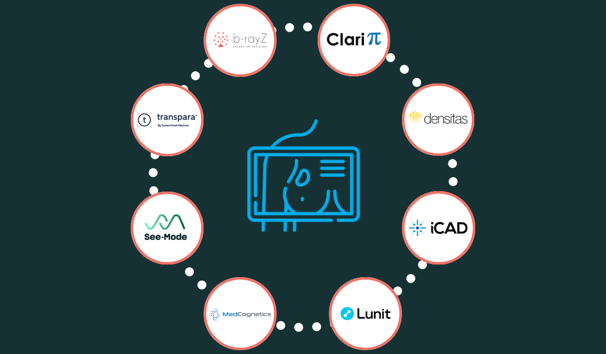
“The importance of breast density measurement cannot be emphasized enough,” said Mina Park McCray, Global Sales and Marketing Manager from AI software company ClariPi, which offers a breast density analysis tool called ClariSIGMAM. “According to the FDA, approximately half of women over the age of 40 have dense breast tissue, which can make cancer detection more difficult, but traditional methods of measuring breast density are subjective and can be prone to human error.”
ClariSIGMAM calculates the percentage of breast density defined as the ratio of fibroglandular tissue to total breast area estimates. The tool uses this numerical value to provide breast density group information according to the BI-RADS categories.
Breast cancer AI specialist iCAD provides a Density Assessment tool that simplifies and standardizes breast density reporting and stratification.
“We know today that density can mask some cancers on mammography, and moreover this is a factor of risk, so it's very important to know a woman's breast density,” said Michele Debain, SVP and General Manager Europe, Middle East, Africa, and Asia Pacific at iCAD. “Our Density Assessment product is a two-part density algorithm, which not only tells you the quantity of dense tissue, but the location of that dense tissue. This also provides a way for departments to standardize their density reporting, because radiologists often don't agree on density scoring.”
With a focus on improving the quality of breast density reporting, AI firm Densitas offers a breast density algorithm called intelliMammo® densityAI™. The algorithm has been clinically validated and has shown that the sensitivity, interval cancer rate, and screen detected cancer rate all poorer in high density breasts compared to low density breasts.
“Our solution addresses the issue of subjective and inconsistent breast density assessment, providing a standardized and automated approach that ensures uniform reporting across all patients – eliminating the variability associated with human subjectivity,” said Jessie Allen, Senior Product Manager at Densitas.
In addition, with b-box plus, a suite of AI tools spanning the breast imaging diagnostic journey, b-rayZ offers the b-density module, which provides real-time and standardized classification of breast density (currently CE-marked only with expecting FDA approval by early 2025), using a 4-point Likert according to the ACR Bi-RADS Atlas 5th Edition and providing standardized recommendations for supplemental ultrasound or MRI examinations.
Korean AI provider Lunit’s MMG solution aims to improve diagnostic accuracy especially for dense and fatty breasts. Their density assessment feature is currently CE-marked.
AI for breast cancer detection
Of course, the role of AI in breast health is already established for computer-aided detection (CAD) tools for both 2D mammography, digital breast tomosynthesis (DBT) and ultrasound, which are increasingly being used to improve cancer detection rates.
The accuracy of early detection of breast cancer previously ranged between 70% and 90%, and CAD applications were developed to reduce the rate of false negatives, typically arising from misinterpretation or oversight of abnormalities. Utilizing pattern recognition algorithms, CAD solutions automatically highlight suspicious areas on mammograms to the radiologist.
CAD algorithms have been found to increase the early detection rates of certain types of breast cancer, particularly those that are more subtle and easily overlooked. [2]
The ProFound AI Breast Health Suite from iCAD has been shown to improve sensitivity and specificity, leading to higher detection rates and fewer false positives.
“Our detection tool works with both 2D and 3D mammography, analyzing every single slice in a 3D or 2D mammogram for suspicious lesions, circling them and giving both a lesion score and a case score,” said Michele Debain from iCAD. It’s almost like having a colleague looking over their shoulder confirming that there's nothing there or there is something that should be looked at more closely.”
Transpara from ScreenPoint Medical enhances the reading of 2D and 3D mammography exams, enabling breast cancer to be detected as early as possible, but also to rule it out.
“Mammography is a screening tool, and 7 out of 8 women screened will not have breast cancer, so it’s important that we confidently and efficiently detect breast cancer, but also confidently say when there is no breast cancer,” said Sanmitra Iwanski, VP of Marketing at ScreenPoint Medical. “The largest screening program in Denmark used Transpara to reduce false positives, while simultaneously reducing radiologists’ workload by up to 62.6%.”
Lunit has developed a CAD tool called INSIGHT MMG, which accurately detects suspicious lesions in mammograms, and the company says that AI can play a key role in supporting radiology departments with limited breast imaging resources.
“A study showed that when a general radiologist uses our product, their detection rate rose by 10%, to 87% matching the level of a dedicated breast specialist,” said Chris McKinney, Sales Director - North America at Lunit. “Not every institution has the luxury of having many breast specialists on staff, so we see this as a way to help all radiologists improve their detection rate. We’ve also seen that our AI is significantly better at detecting stage T1 and in stage negative cancers, which are the types of cancers that really give radiologists problems.”
Lunit’s solution for digital breast tomosynthesis, INSIGHT DBT, is an AI solution that identifies and classifies suspected areas for breast cancer. The solution automatically analyzes 3D images and provides visualization and quantitative estimation of the presence of malignant lesions.
While mammography is the most common modality associated with CAD in breast health, ultrasound is also an area to benefit from the use of AI. Australian company See-Mode has developed a tool that automatically reads and assesses ultrasound images, detecting lesions and assigning feature classifications in line with the BI-RADS reporting scheme, producing a clinical report for the radiologist to complete.
“See-Mode leverages AI to automatically generate Sonographer worksheets, saving them considerable time while also eliminating data entry errors and other inaccuracies. See-Mode additionally sends study findings and preliminary impressions directly to Radiologist report templates, reducing dictation time,” said Shehan Bala, Head of Product at See-Mode. “In the near term, workflow improvement is where we see AI being most impactful - taking care of things that Sonographers and Radiologists find tedious, enabling them to focus on higher value activities while also decreasing burnout.
Some CAD tools can also prioritize cases, helping radiologists focus on studies that have a higher degree of suspicion. There are also standalone solutions that can help with study prioritization. AI developer MedCognetics has developed a tool called CogNet AI-MT, which alerts radiologists about mammograms containing at least one suspicious finding. The suspicious study indication enables worklist prioritization and optimization based on clinically relevant information.
“We are proud to highlight that our solution, CogNet AI-MT, not only optimizes worklist prioritization but also sets a new standard in Unbiased AI,” said Ron Nag, CEO at MedCognetics. “Trained on a diverse patient population, CogNet AI-MT performs reliably across all patient types, ensuring equitable and accurate diagnostic support. This commitment to inclusivity and precision empowers radiologists to deliver superior patient care with greater efficiency and confidence."
Evaluating breast cancer risk with AI
Increasingly, the use of AI is expanding from identification of potentially suspicious areas in a mammogram towards assessing a patient’s risk of breast cancer. This information can be used to enable more personalized screening plans, helping to detect cancer earlier. Lifetime breast cancer risk estimates using risk models such as the Tyrer-Cuzick risk model now incorporate breast density as a critical risk factor.
AI company iCAD’s ProFound AI Risk tool is an image-based breast cancer risk estimation algorithm. By evaluating several data points in the patient and scan image, it calculates a short-term risk score of developing breast cancer over the next one or two years. Profound AI Risk has been shown to be more than twice as sensitive as established methods of identifying women at high risk of developing breast cancer in one to two years, and also predicts the level of aggressiveness of the future cancer.
“Our risk analysis tool can evaluate a woman's risk of developing breast cancer in the short term, which is really exciting,” said Michele Debain at iCAD. “Combining AI with mammographic features and the age of a woman, we can accurately determine their risk of developing cancer in the next one to two years. Then they can decide with their radiologists if they should come in more often to have a mammogram or perhaps do their MRI or an ultrasound.”
In addition, ScreenPoint Medical’s Transpara solution includes an Exam Score feature, a tool that categorizes exams using either a 3-point scale (Elevated, Intermediate, Low) or a 10-point scale for an overview of the level of suspicion. The higher the score, the higher the risk of cancer in the mammogram.
A clinical study in Sweden investigated use of Transpara’s Exam Score as an alternative to double reading, with intermediate or low risk mammograms (90% of all studies) read by only one reader while the 10% labelled as in the highest numeric risk category were read by two readers.
“Use of the Transpara Exam Score in the randomized controlled trial showed that the workflow helps detect more cancers while safely reducing workload by 44% with no change in recall rate,” said Sanmitra Iwanski from ScreenPoint Medical. “Clinical research shows that low risk Transpara exam scores have a 99.97% negative predictive value.”
The role of AI in image quality
The Mammography Quality Standards Act (MQSA) dictates the standards required to ensure that all women have access to quality mammography services. Clinical image quality is a key aspect of the MQSA Enhancing Quality Using the Inspection Program (EQUIP) compliance. AI tools are now helping radiology centers minimize the administrative burden associated with ensuring compliance with MQSA EQUIP standards.
The quality of clinical images in mammography is crucial for accurate breast cancer detection. Inadequate positioning and poor image quality can significantly reduce the sensitivity and specificity of mammography, leading to misdiagnosis and unnecessary follow-ups.
Through its intelliMammo® intelliPGMI™ and Navigator™ solutions, Densitas also provides standardized image quality assessment of mammograms, flagging poor clinical image quality, benchmarking performance, and tracking corrective actions.
“Maintaining mammography quality is not just a matter of meeting compliance requirements, it’s a matter of equity and access to all women, not just a small random sample whose mammograms are reviewed in prep for an inspection. Every woman deserves to have her mammogram evaluated for optimal clinical image quality every time she has a mammogram taken.” said Mo Abdolell, CEO of Densitas.
Beyond its diagnostic imaging tools, b-rayZ's b-box plus also offers the b-quality module (CE and FDA cleared) for image quality assessment. The b-quality module provides real-time information on the quality of mammograms, helping technicians correct and avoid errors in the positioning of patients to reduce patient recalls. The b-smart dashboard (CE and FDA cleared) allows to monitor the performance of the breast unit(s), leading to lean management of resources and optimization of procedures. b-smart also offers the option to generate automatic quality reports to the authorities.
“We provide real time information on the PGMI-based mammography quality assessment that allow technicians to correct recurrent errors in the positioning of the patients in the mammography scanner through graphical feedback,” said Alex Ciritsis, CTO at b-rayZ. “We also help identify areas for improvement that consultants can use to deliver targeted training.”
Challenges of AI adoption in breast imaging
In the evolving field of breast imaging, the integration of AI clearly offers hospitals and imaging centers a promising pathway towards enhanced patient care, increased efficiency, and streamlined operations. However, the challenge lies in navigating the multitude of AI solutions available and determining which one best suit the specific needs of a healthcare provider.
The rapid growth of AI tools in mammography has resulted in the development of numerous algorithms, many of which seem quite similar. The primary challenge for healthcare providers is to determine which AI application works best in their specific context. This decision can be influenced by various factors, including the patient population, the type of imaging equipment used, and other institutional specifics. Partnering with a single AI platform provider offers a strategic solution to these challenges.
The benefits of a platform approach
Adopting a platform like Blackford allows healthcare organizations to test different AI solutions using their own data before making a decision. This enables providers to conduct retrospective analyses on the platform, assessing which application performs best in their environment. By offering multiple applications for critical use cases such as breast density assessment, providers can ensure they select the most effective tools for their needs.
While each AI algorithm may offer slightly different features, a platform approach essentially provides centers with a comprehensive suite of mammography tools. This ensures the most extensive coverage of available tools, as no single vendor offers a complete solution for every use case. By centralizing access to various AI solutions through a dedicated platform, healthcare providers can efficiently evaluate, implement, and maintain a diverse array of imaging AI applications. This not only maximizes resource utilization but also reduces deployment timelines and financial overheads.
A platform-centric approach simplifies several processes by consolidating contact management, deployment procedures, and ongoing support into a single interface. Platforms seamlessly integrate with existing hospital systems, ensuring smooth performance, particularly in handling Digital Imaging and Communications in Medicine (DICOM) interfaces. This integration is crucial for maintaining operational efficiency and ensuring that new AI tools can be adopted without disrupting existing workflows.
Stay on top of the latest developments
Platforms also help alleviate the issue of analysis paralysis by providing access to a portfolio of pre-vetted AI applications across various medical disciplines beyond breast imaging, including cardiology, neurology, pulmonology, trauma, and MSK. By offering comprehensive bundles of AI applications through a single contract, platforms eliminate the complexities associated with acquiring individual algorithms. This simplifies the procurement process and ensures that healthcare providers have access to a broad spectrum of AI solutions.
Ultimately, embracing a single-platform strategy enables breast health clinicians and their institutions to access a diverse array of AI solutions, facilitating the swift adoption of new technologies and the evaluation of applications tailored to their specific needs, and the needs of their patients. This approach not only improves patient care and operational efficiency but also ensures that healthcare providers are at the forefront of any new technological advancements in breast imaging AI.
[1] Journal of Breast Imaging, Volume 5, Issue 6, November-December 2023
[2] Clinical Imaging, Volume 52, 2018
If you want to learn more about our partners and their thoughts on this topic, we have a webinar coming out soon!


